Loading...
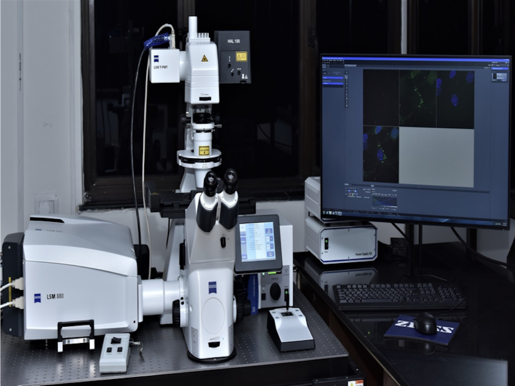
Laser Scanning Confocal Microscope's Details

Equipment Name :
Laser Scanning Confocal Microscope
Make :
Carl Zeiss
Model :
LSM 880
Status :
 Functional
Purchase Date :
26 Dec 2017
Warranty/AMS :
Warranty CAMC
Unit of Use :
Per 2 Hour
Consumables/Services Provided :
No
Prior discussions or payment required? :
Yes
Technical Help Provided :
Assist
Minimum Booking per unit of Use :
-
Maximum Booking per unit of Use :
-
Contact Person :
Prof. Deepak T. Nair
Email :
Phone No. :
-
Technical Personnel :
Damini
Email :
Phone No. :
0129-2848619
Location :
ATPC Optical Microscopy Facility
Working Hours
The coherent light (excitation laser) passes through a pinhole aperture that is situated in a conjugate plane (confocal) with a scanning point on the specimen and a second pinhole aperture positioned in front of the detector (a photomultiplier tube). As the laser is reflected by a dichromatic mirror and scanned across the specimen in a defined focal plane, the emission from points on the specimen (in the same focal plane) pass back through the dichromatic mirror and are focused as a confocal point at the detector pinhole aperture. Due to this aperture, the extraneous light (out of focus light) is not detected by the detector thereby resulting in sharp images.
About Microscope: The Microscope consists of an inverted fluorescence research microscope (Axio observer 7) with following objectives:
10X Plan Apochromat (NA 0.45)
40X Plan-Apochromat Oil immersion DIC (NA 1.30)
63X Plan-Apochromat Oil immersion DIC (NA 1.40)
Observation and imaging units: A metal halide illuminator (HXP120) and HAL100 illuminator for sample observation and 5 lasers including Multi Argon lines for confocal imaging.
Diode laser 405 nm
Laser Argon Multiline 458/488/514 nm
Laser HeNe 543 nm
Laser HeNe 594 nm
Laser 633nm
Detection unit:
Transmitted light detector T-PMT
LSM 880 confocal scan head with 2MA-PMT and 1 GaAsP (for spectral detection)
High resolution digital microscopy camera AxioCam 503 mono


