Loading...
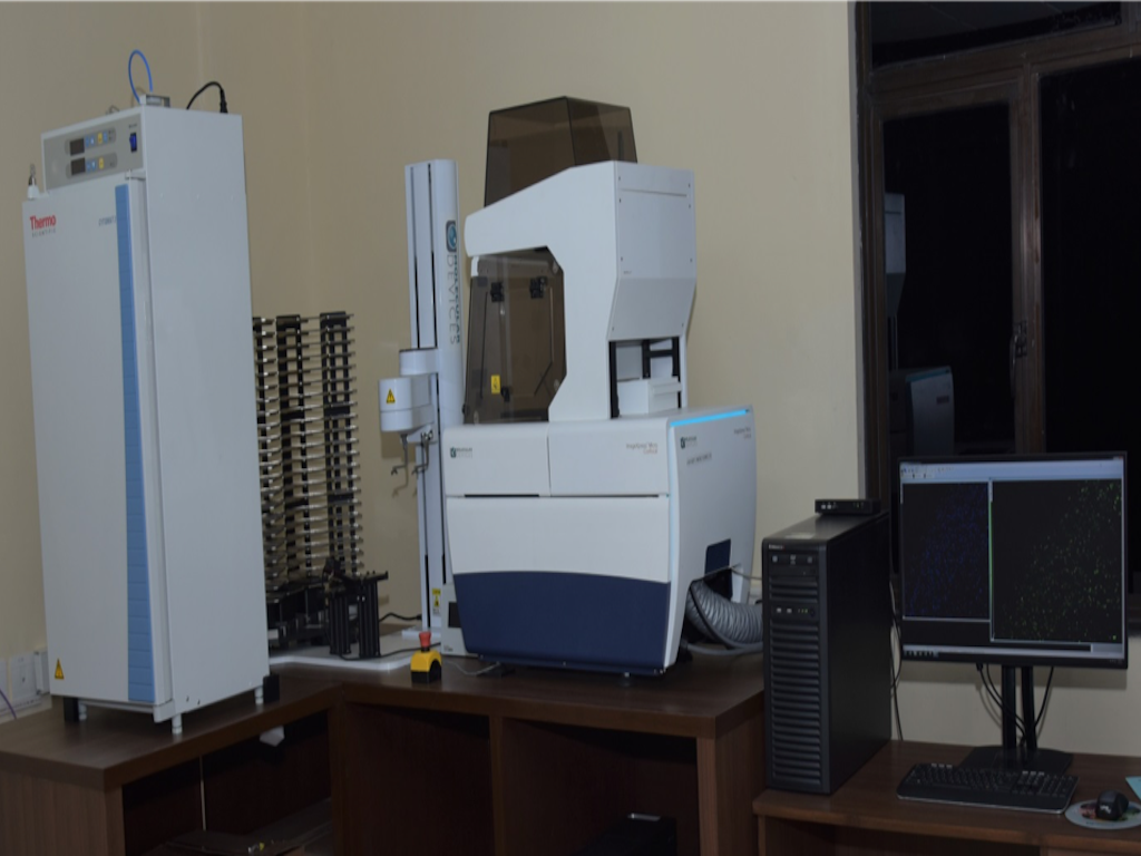
Micro Confocal High Content Imaging System's Details

Equipment Name :
Micro Confocal High Content Imaging System
Make :
Molecular Devices
Model :
ImageXpress
Status :
 Functional
Purchase Date :
24 Nov 2017
Warranty/AMS :
AMC
Unit of Use :
Per 2 Hour
Consumables/Services Provided :
No
Prior discussions or payment required? :
Yes
Technical Help Provided :
Assist
Minimum Booking per unit of Use :
-
Maximum Booking per unit of Use :
-
Contact Person :
Prof. Deepak T. Nair
Email :
Phone No. :
-
Technical Personnel :
Damini
Email :
Phone No. :
0129-2848619
Location :
ATPC Optical Microscopy Facility
Working Hours
The ImageXpress system is an integrated cellular imaging and analysis system that is designed for rapid, automated screening of fluorescently labelled fixed and live cells in microplates. It also provides environmental control for live cell imaging, moves fluids to and from cells for compound addition. The ImageXpress Confocal system is a high-content imaging system that can switch between widefield imaging and confocal imaging. The rapid autofocus and precision sample movement features of the microscope allow large numbers of high-resolution images to be acquired in the shortest possible time. All key optical and mechanical elements are motorized, with asynchronous command execution, allowing complete real-time control of the instrument configuration through the MetaXpress high-content image acquisition and analysis software. When used in combination with the powerful image analysis capabilities of the MetaXpress software, the instrument becomes an extremely flexible and programmable device, ideally suited for user-defined, high-speed automated assays.
Instrument Specifications
ImageXpress Micro Confocal System is a fully automated confocal microscope with pinhole sizes of 50 µm and 60 µm.
It is capable of taking images from a microplate (96 well/ 384 well / 1536 well) or from a microscopic slide.
It is capable of detecting and imaging multiple fluorescent signals simultaneously.
Transmitted light imaging can be done for non-fluorescent cells.
Z-stack images can be acquired and analysed.
Environmental control- multi-day, live cell time-lapse imaging under appropriate temperature, humidity and atmospheric conditions can be performed.
This system also contains single-channel, fluidics robot. This robot picks up tips from a tip box and draws fluid from and delivers fluid to a given well on the compound and sample plates. The robot is capable of dispensing volumes between 3 μL and 200 μL.
This system also has a hotel shelf with capacity for 88 standard microplates, where plates can be stacked for imaging. With the help of Sample Handling Robotic Arm, the plates can be removed from hotel shelf and can be loaded onto ImageXpress Micro Confocal System where images are acquired, and then returned to a home location.
This entire system has been integrated with Cytomat 2C automated incubator through a Sample Handling Robotic Arm so that multiple plates can be scanned for time-lapse live cell imaging. This automated incubator has a capacity for 42 standard microplates.
Automated barcode reader is also a part of this system.
Light Source
The external light source for the ImageXpress Micro Confocal System is a solid-state light source that has a rated lifetime of more than 20,000 hours. It has light source for excitation of DAPI, FITC, TEXAS RED, Cy3, Cy5 wavelengths.
Objectives
4X Plan Apo λ NA 0.20
10X Plan Apo NA 0.45
20X S Plan Fluor ELWD NA 0.45
40X S Plan Fluor ELWD NA 0.60
60X S Plan Fluor ELWD NA 0.70
100X CFI L Plan EPI NA 0.85
Applications
Application modules are the foundation of the MetaXpress Software analysis workflow. These modules provide automated image analysis for biology-specific applications, interactive image segmentation, and data results, and options for data recording and management. Currently, Our MetaXpress software has the following modules:
Angiogenesis Tube Formation
Cell Cycle
Cell Health
Cell Proliferation
Cell Scoring
Live Dead
Micronuclei
Multi Wavelength Scoring
Translocation
Neurite Outgrowth


